

Home
Product Details
Unity
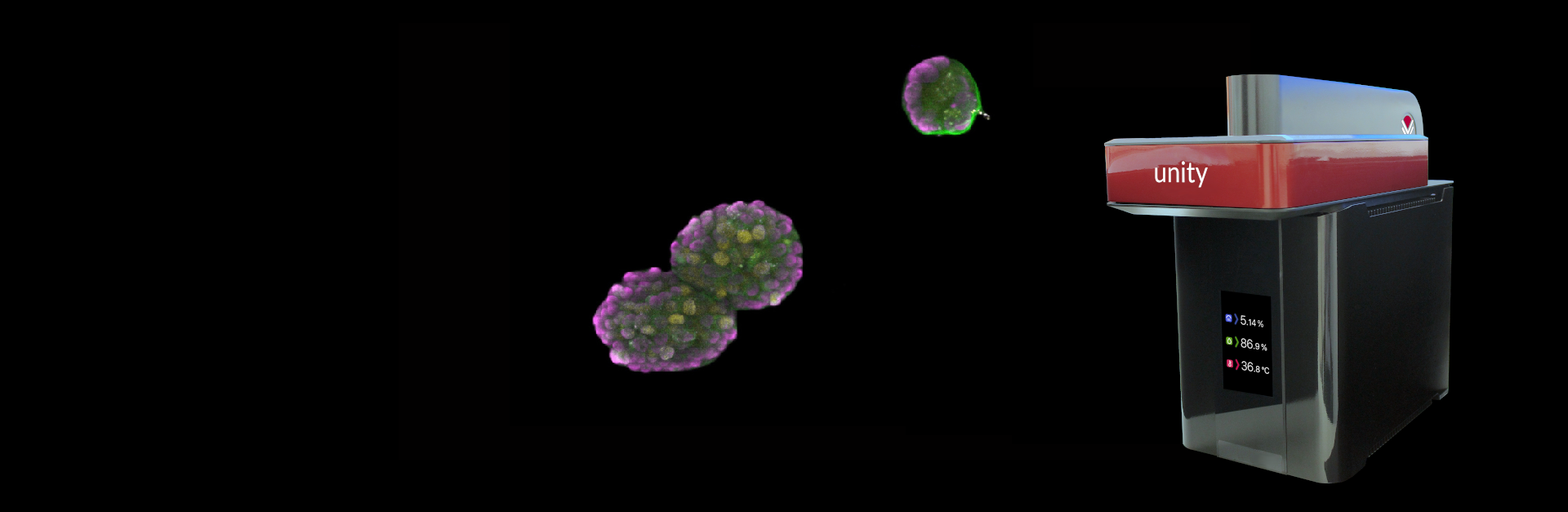
The All-In-One Benchtop Confocal

Complete Confocal Microscope for Live Cell Imaging
Introducing Unity, a new breed of user friendly bench-top laser-free confocal microscope bringing affordable high speed, high resolution imaging to your laboratory.
Flexible and Adaptable
Unity for all your imaging experiments:
TIME-LAPSE
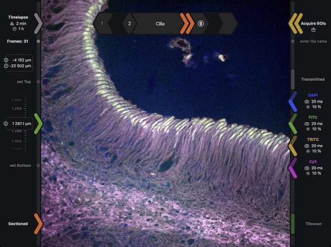
Long Time-Lapse Experiments
Time-lapse is easy with Unity.
Simply define your region of interest.
Then it’s:
With its go anywhere design and integrated vibration dampening, Unity is ideal for use in the light and airy open laboratory.
Unity for live-cell imaging
The CAT-3 ready Unity
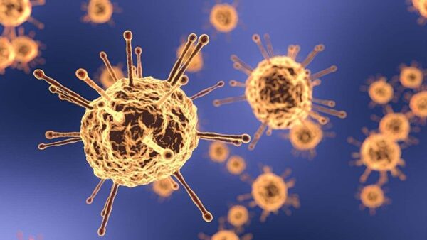
The compact and fully integrated design of Unity makes it the ideal confocal microcope for use in the CAT-3 laboratory.
Unity is extremely easy to use with minimal interaction and it’s construction allows for the use of typical decontamination procedures.
TILESCAN
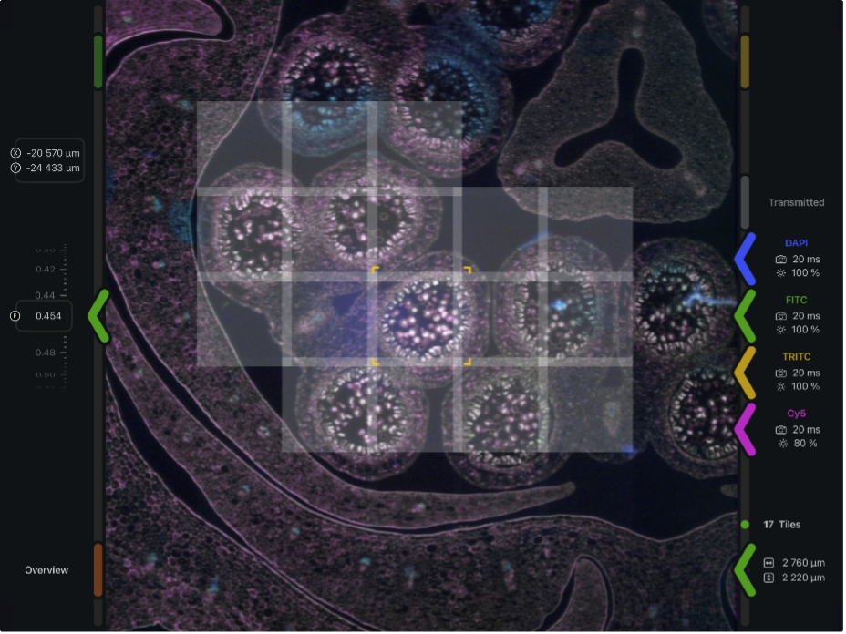
Drag-Out a Tilescan Area
Drag out a wire frame over the desired tilescan area to define tiled acquisition protocol. Tap on individual tiles to de-select them, if a non-rectangular acquisition area is desired.
Unity does the rest.
Tile Stitching
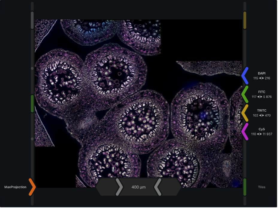
High-fidelity stitched images are generated by Unity for image data review purposes. These images are stored as pyramidal multi-resolution files, thus enabling display at high zoom settings without any loss of on-screen resolution.
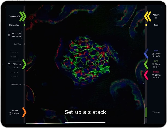
Z-STACK
FAST MULTI-CHANNEL
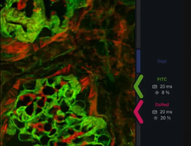
WIDEFIELD / CONFOCAL
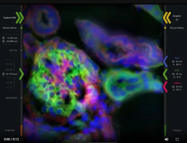
MULTI-POSITION
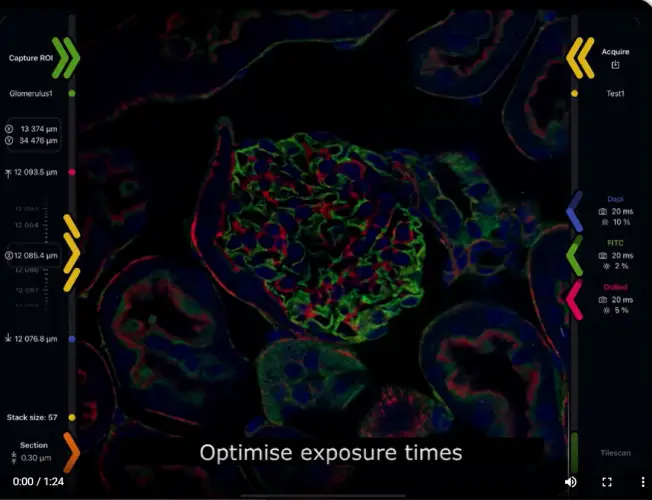
AUTOFOCUS
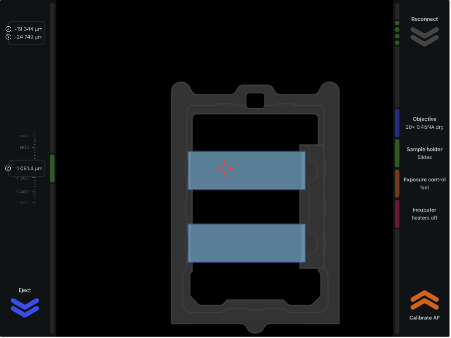
IR autofocus
Unity is equipped with hardware autofocus, which uses infrared LED reflected from the coverslip to measure and maintain the stable focus position when sample is moving or during extended time-lapse aquisitions.
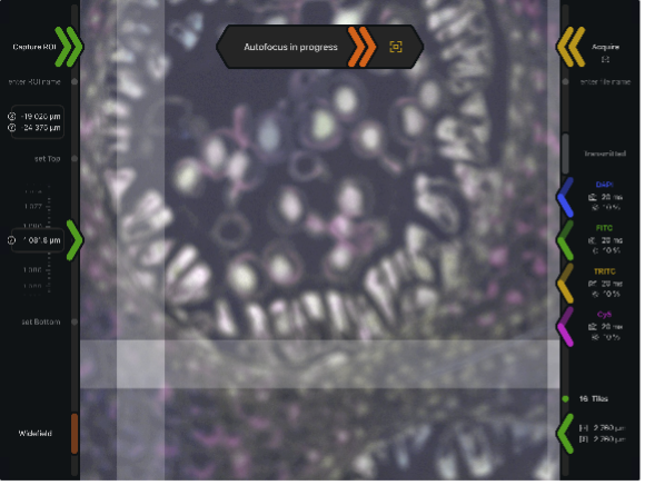
Software autofocus
Unity also has image-based software autofocus. In this mode z-position is adjusted for the best quality, sharpest image. Software autofocus is particularly useful in the situations where the sample position can't be reliably deduced from the coverslip. It can be used on its own or together with IR autofocus.
PLATES, DISHES AND SLIDES
Plates, Dishes and Slides with Unity Sample Holders
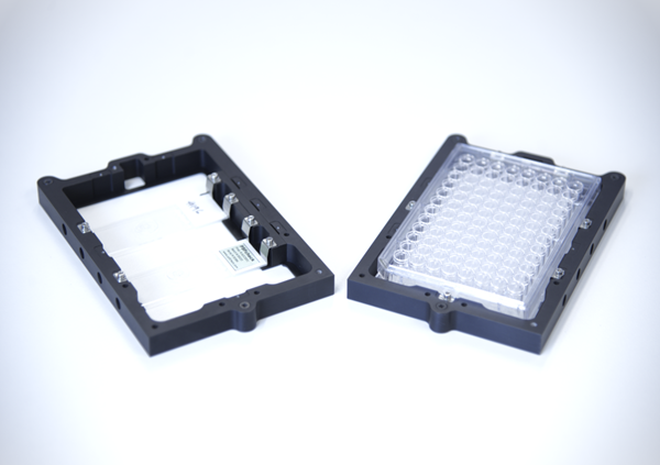
Unity can be supplied with three interchangeable sample holders allowing samples to be imaged, whether they are mounted in multi-well plates, petri-dishes or slides.
Each of the Unity sample holders has a retention system to hold the sample securely and a magnetic locating system which connects to Unity’s XY stage in the holder.This system ensures the sample holder position is securely held throughout imaging experiments.
Imaging multi-well plates
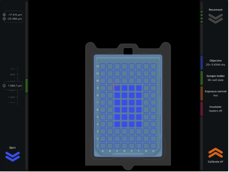
Unity software has convenient tools that simplify imaging of samples contained in multi-well plates. Wells can be selected for acquisition individually or by blocks, each well being assigned a labelled region of interest with an automatically generated acquisition protocol. The parameters of these protocols are individually adjustable and IR autofocus functionality is available to maintain focus over long timelapses
ENVIRONMENTAL CONTROL
Unity's live cell capability
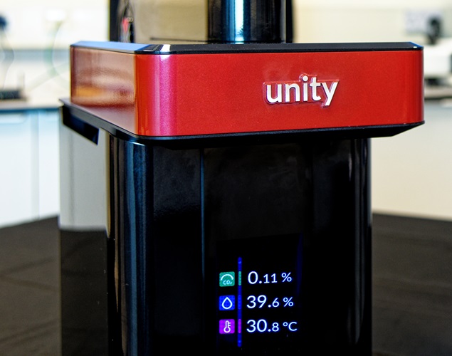
Unity’s red cased incubator sample stage provides heating and CO2 gas connections for sample environment control, allowing your live-cell samples to be maintained at 25-370C and under CO2 for the duration of your longer term imaging experiments (external humidifier and CO2 mixer required).
SOFTWARE
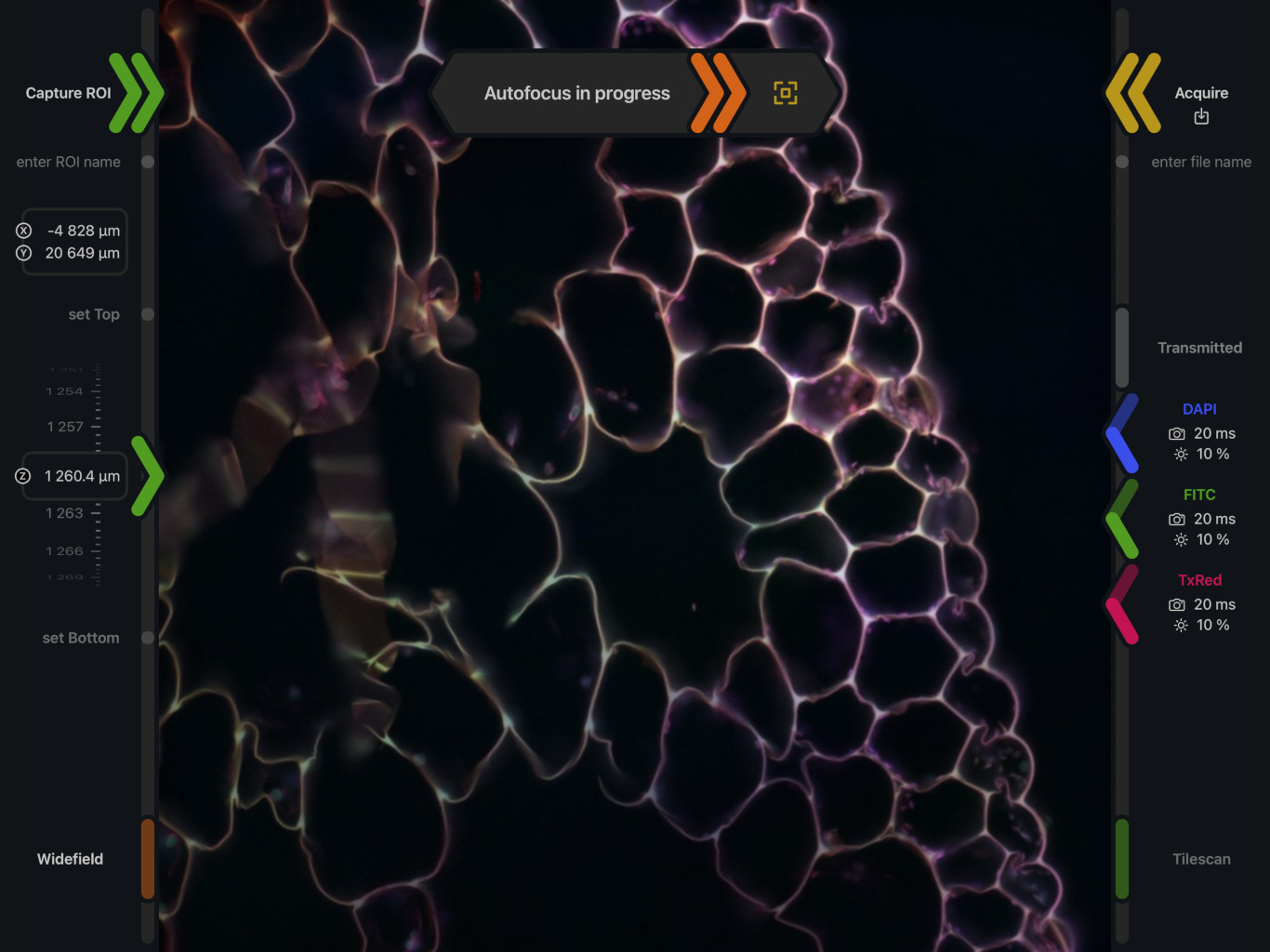
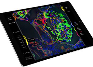
Tap, Drag and Swipe with Unity’s iOS Software
Unity iOS provides all the functionality required for the set-up, control and review of confocal and widefield imaging experiments in a quick and easy to use iPad application.
Imaging is easy with Unity.
Simply:
Unity's iPad Controller
Unity comes complete with an Apple iPad tablet computer for full fingertip control.
The iPad communicates directly with Unity over a dedicated and secure private wireless connection, issuing commands and visualising data saved on Unity’s built in image server.
To increase the speed of the data stream response, images are optimised for faster feedback and display on the iPad. The full high resolution collected images are permanently stored on Unity’s internal server.
The iPad works as a remote control system with all commands controlled by Unity’s software and firmware.
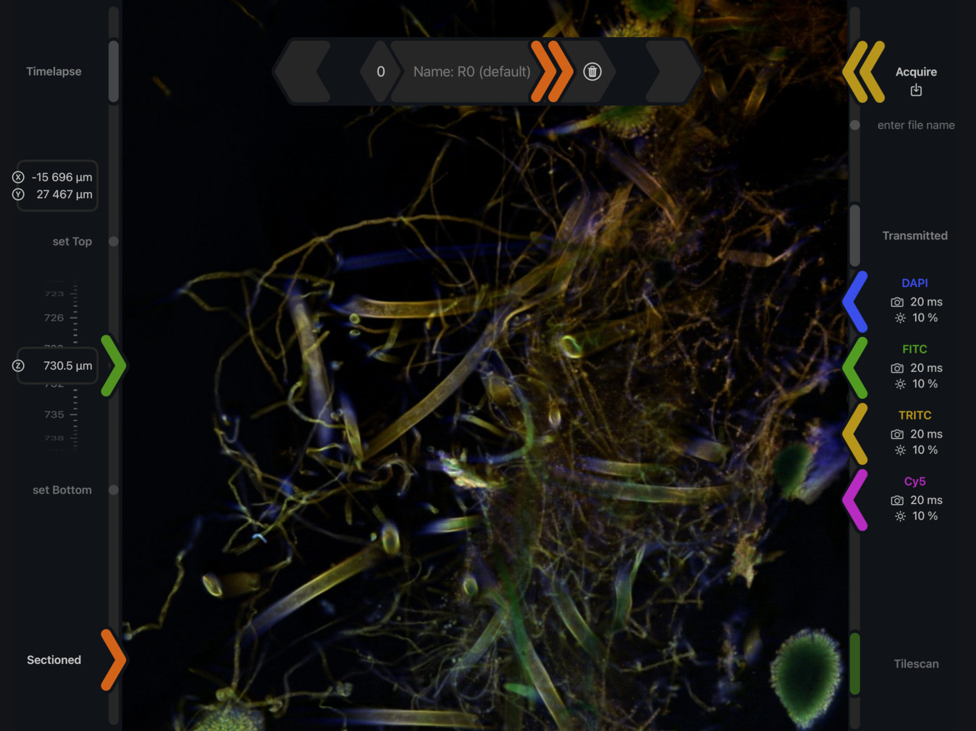
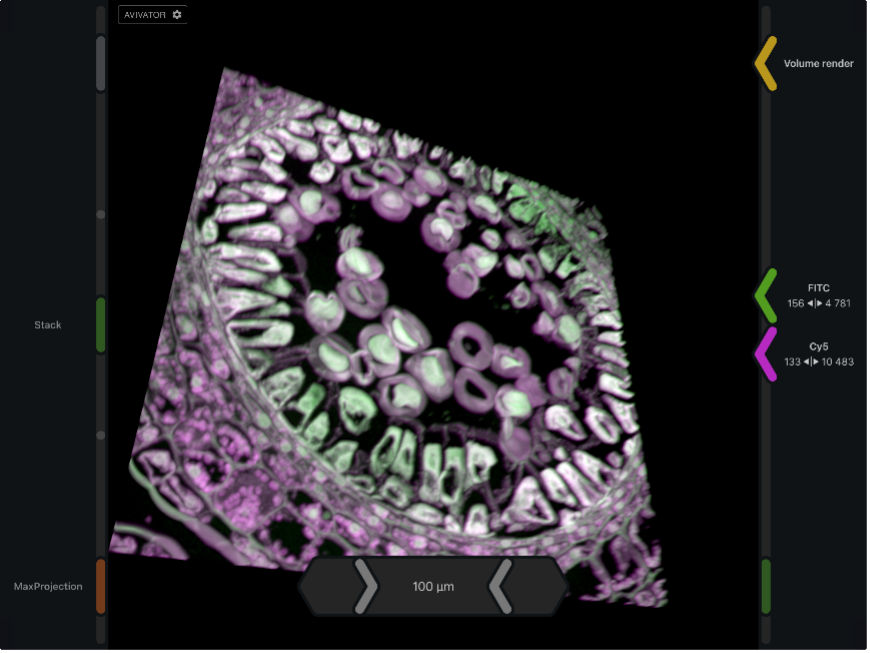
Image Data Review
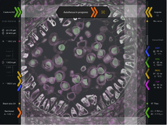
Unity offers extensive image data review capabilities, including z-stack browsing, timelapse movies, tile stitching and 3D rendering, as well as full control over fluorescence channel colours and contrast ranges.
LIGHTING
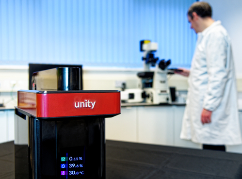
Unity's Choice of Light
Our patented confocal technology is significantly more light efficient, meaning Lasers are not needed.
Instead, Unity uses our in-house powerful multispectral LED source making Unity:
CAMERAS
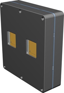
Unity's 2x4 Megapixel Scientific CMOS Camera
At the heart of Unity is a custom designed large format and high performance sCMOS digital camera. Developed in collaboration with Ximea, the camera has been designed specifically for use in Unity and for confocal imaging experiments.
Unity’s camera has a novel design, consisting of two 2k x 2k scientific grade CMOS chips mounted side by side with shared electronics such that the two chips are pixel to pixel registered allowing Aurox’s patented approach to laser-free confocal imaging.
Unity uses the camera to record both confocal and widefield images in both the low magnification sample overview mode (large field of view of 4mm x 4mm) and also in the high magnification 4-100x mode for the imaging of high resolution Z-stacks.
SYSTEM HARDWARE
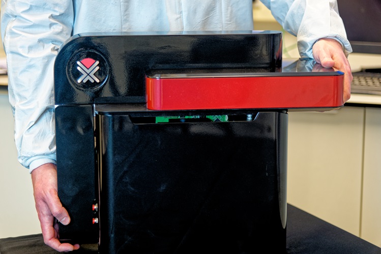
Plug and Play
Designed for the laboratory benchtop, Unity is extremely compact and self-contained, weighing less than 25 Kg. Gone are the tangle of cables found on other confocal systems. Unity requires a single power cable and has rear ports for the connection of a CO2 supply. Unity can be networked wirelessly, but it also has a wired network connection and multiple USB3/USB-C ports if required.
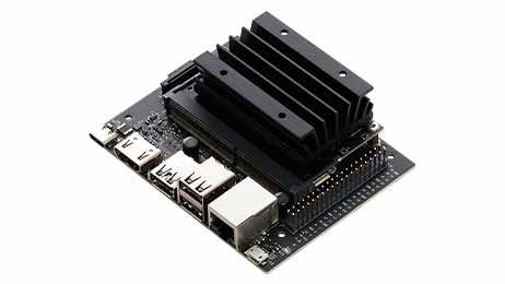
Unity's CPU and Image Server
Unity does more than just collect high quality confocal image data. Unity also stores your data on a 4TB SSD integrated drive and makes your data accessible, wherever you are, over a secure network.
REMOTE ACCESS
Remote Access
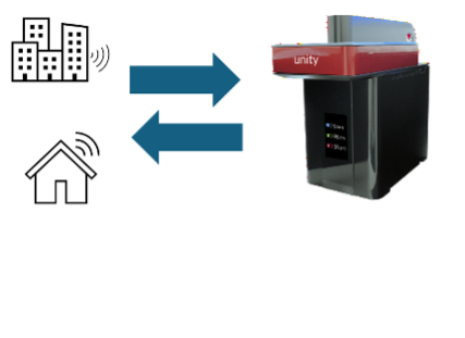
Remote access allows control of experiments, inspection of the acquired data, training and support from anywhere in the world. Unity offers remarkable networking flexibility and is able to interact directly with local Network Attached Storage or Cloud Data Storage solutions.
OPTICAL PATH
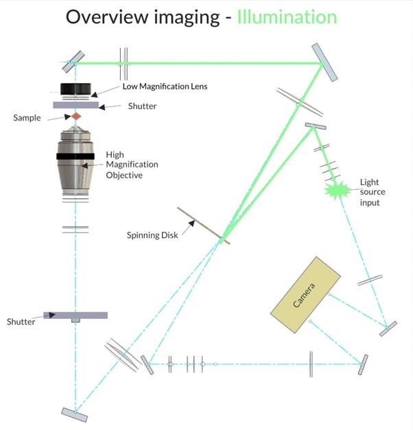
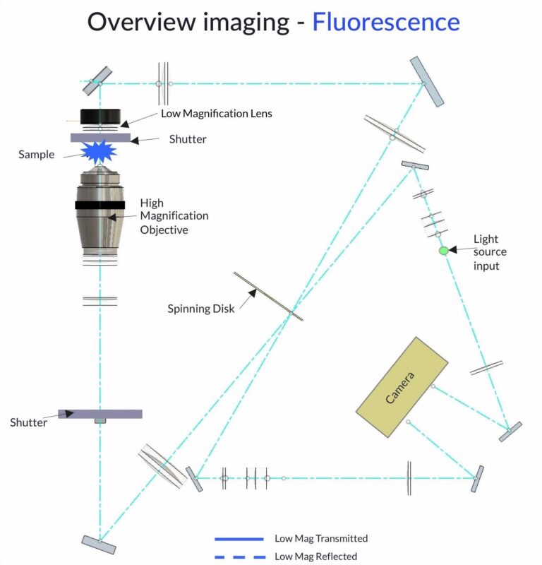
Unity's Patented Optical Path
The patented optical path is central to the design of Unity and its enabling technology. It allows sample imaging from both above and below the sample, providing confocal and widefield imaging whilst maintaining an extremely compact size.
Scroll through the images to see this in action.
SPECIFICATIONS
| Key specifications | Unity |
|---|---|
| Resolution (lateral × axial) | 275 x 700 nm with 1.45 N.A oil-immersion objective |
|
Confocality |
0.9 μm with 100x 1.45 N.A oil-immersion objective |
| Disk | single-sector structured illumination disk. |
| Minimum exposure | 20 ms |
| Maximum frame rate | Up to 22 fps (3 colour) |
| Up to 40 fps (1-colour) | |
| Imaging channels | 4 integrated filter channels. |
| Channel switching | < 1ms. |
| Detector | Integrated sCMOS 2× (2048 x 2048), 6.5 µm pixels. |
| Excitation range | 370 - 700 nm |
| Emission range | 410 - 750 nm |
